Traditional 2D Dental X-rays VS 3D Cone Beam X-rays
January 10th, 2024 | 4 min read

In the ever-evolving field of dentistry, imaging technology plays a crucial role in diagnostic and treatment planning. Traditional dental X-rays and 3D Cone Beam X-rays are two key modalities, each with unique advantages and specific applications.
From our team at NYC Smile Denign’s experience in the dental field, we’ve seen firsthand how the right imaging choice can significantly impact both the diagnosis and the success of treatment plans.
This article focuses on these two technologies, helping you understand their differences and guiding you in making informed decisions about your dental care.
Understanding Traditional Dental X-rays
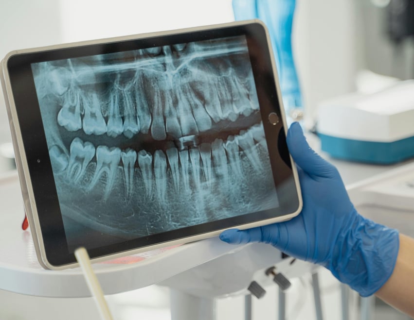
Traditional dental X-rays, also known as 2D X-rays, have been the cornerstone of dental imaging for decades. They provide a flat, two-dimensional view of the teeth, bones, and hard tissues of the mouth.

Key Advantages of Traditional 2D X-rays
One of the most notable advantages of traditional X-rays is their ability to capture high-resolution 2D images. This level of detail is crucial for accurately detecting cavities, particularly those that are not visible to the naked eye. Additionally, they are excellent for examining the condition of tooth roots, identifying any issues that may lie beneath the gum line, and assessing the bone levels around the teeth. This precision is essential for diagnosing various oral health conditions and planning appropriate treatments.
Traditional 2D X-rays are a staple in most dental practices due to their ease of use, instant processing time, and reduced radiation. This accessibility makes them an ideal choice for routine dental check-ups, as they can be performed rapidly and provide immediate results. This speed is particularly beneficial in emergency dental situations where a swift diagnosis is necessary.
Ideal Scenarios for Use
Traditional dental X-rays hold a place of significant importance in various dental scenarios due to their precision and reliability. They are particularly useful in routine dental examinations, where a comprehensive view of the patient’s oral health is required. These X-rays allow dentists to monitor the ongoing condition of teeth and gums, ensuring any changes or developments are noted and addressed promptly.
In the realm of cavity detection, traditional X-rays excel. They are adept at revealing the early onset of tooth decay, often detecting cavities before they become visible or symptomatic. This early detection is crucial in preventing the progression of decay, which can lead to more severe dental issues if left untreated.
For periodontal disease assessment, traditional X-rays are invaluable. They provide essential information about the level of bone surrounding the teeth, an important factor in diagnosing and monitoring gum disease. Through these X-rays, dentists can assess the severity of the periodontal condition, track its progression, and plan effective treatment strategies.
Additionally, traditional X-rays play a pivotal role in the preliminary evaluation phase of various dental procedures. Before undertaking treatments such as dental implants, root canal therapy, or complex extractions, a detailed understanding of the underlying dental structure is essential. Traditional X-rays offer this insight, allowing dentists to plan procedures with a higher degree of accuracy and safety, ensuring better outcomes for patients.
Exploring 3D Cone Beam X-rays
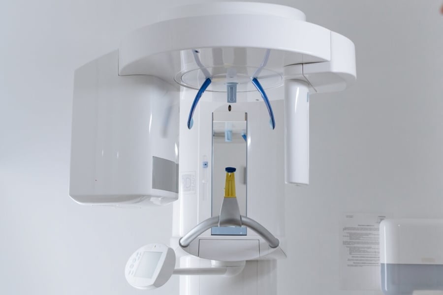
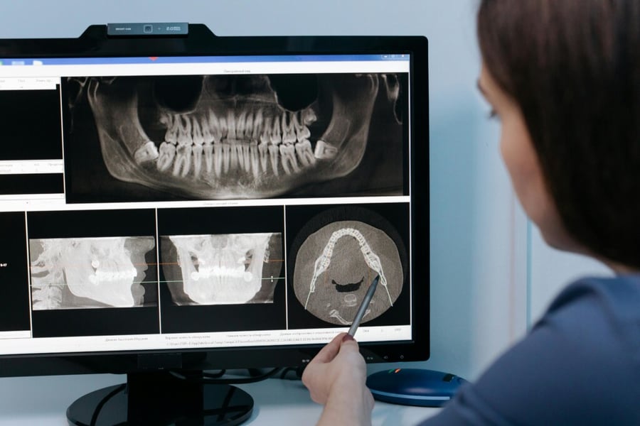
3D Cone Beam X-rays, or Cone Beam Computed Tomography (CBCT), provide three-dimensional images of the teeth, soft tissues, nerve pathways, and bones in a single scan.

Key Advantages of 3D Cone Beam X-rays
The most striking advantage of 3D Cone Beam X-rays lies in their ability to provide a complete three-dimensional view of the oral and maxillofacial region. This comprehensive perspective is pivotal in understanding the intricate spatial relationships between different structures in the mouth and jaw. Unlike traditional X-rays, which offer a limited two-dimensional view, 3D imaging allows for the visualization of anatomical structures in a more natural and intuitive way. This depth of detail is particularly beneficial when assessing areas that are complex or difficult to view with standard X-rays, such as the sinuses, nasal cavity, and nerve pathways.
3D Cone Beam X-rays are incredibly versatile in both diagnostic and planning phases of dental care. They play an essential role in implant planning, where a detailed understanding of bone structure, density, and the location of critical anatomical features, like nerves and blood vessels, is crucial. This technology ensures that implants are placed precisely and safely, significantly reducing the risk of complications.
Ideal Scenarios for Use
3D Cone Beam X-rays have revolutionized dental imaging, especially in complex dental cases where detailed visualization is crucial. One of the primary applications of this technology is in the planning of dental implants. The 3D imagery provides a comprehensive view of the patient’s jawbone, including bone density and structure, which is essential for determining the optimal placement of implants. This level of detail ensures a higher success rate for implant procedures by allowing for precise surgical planning.
In orthodontic treatments, 3D Cone Beam X-rays offer an unparalleled advantage. They allow orthodontists to view the teeth and supporting structures from various angles, aiding in the creation of more accurate and effective treatment plans. This technology is particularly beneficial in assessing tooth alignment and jaw development, playing a critical role in devising personalized orthodontic strategies.
The evaluation of jaw disorders, such as temporomandibular joint (TMJ) disorders, is another area where 3D Cone Beam X-rays prove invaluable. They provide a clear view of the bones and joints of the jaw, enabling dentists to diagnose and understand the complexities of TMJ disorders more effectively. This comprehensive view assists in developing more targeted treatment plans to alleviate symptoms and address the root cause of the disorder.
Furthermore, 3D Cone Beam X-rays are instrumental in more intricate endodontic cases. They offer detailed images of the tooth’s root structure and canals, which are vital for successful root canal treatments. This technology allows endodontists to navigate the intricate anatomy of a tooth, ensuring thorough cleaning and filling of the root canals, thereby increasing the chances of a successful treatment.
Overall, 3D Cone Beam X-rays are a powerful tool in the dental field, offering detailed and three-dimensional insights that are critical for the successful planning and execution of complex dental procedures. Their ability to provide in-depth and accurate images makes them an indispensable resource for advanced dental diagnostics and treatment planning.

Conclusion: Tailoring Technology to Your Dental Health
Both traditional dental X-rays and 3D Cone Beam X-rays have their distinct places in dental diagnostics and treatment planning. Understanding their differences and applications enables you to engage in more informed discussions with your dental care provider. Whether it’s a routine check-up or a complex dental procedure, choosing the right imaging technique is a step toward ensuring effective and personalized dental care.
Now that you have a clearer understanding of the differences between traditional dental X-rays and 3D Cone Beam X-rays, you might be wondering about the insurance aspects of these procedures. To gain insights into what your dental insurance may cover, we encourage you to read our informative blog, “What You Need to Know About Dental Insurance.” This resource will provide you with valuable information to help navigate insurance coverage for various dental imaging techniques.
If you’re considering which imaging technique is best suited for your dental needs, we invite you to schedule a consultation with us at NYC Smile Design. Our team of experts is equipped to provide you with comprehensive information and guidance, ensuring that you have all the necessary dental records and insights for your treatment plan. Contact us today to explore your options and take a confident step toward optimal dental health.
Topics:

